Hand Surgery Source

DISLOCATION, WRIST RADIOCARPAL

Introduction

Radiocarpal joint dislocations are uncommon, with an incidence of ~0.2% of all wrist injuries.1 They nearly always result from high-energy trauma that causes extremes of hyperextension and pronation, such as a fall from a height or motor vehicle crash and are most frequently seen in young, active men.2 The mechanism of injury is thought to be a severe shear or rotational insult that transmits the force from lateral to medial through the platform of the radius.3 The majority of these dislocations involve associated injuries—particularly radial styloid fractures—with purely ligamentous injuries being extremely rare.4 Conservative treatment may be appropriate for some cases of radiocarpal dislocation with no intracarpal damage, but most surgeons prefer surgical intervention for optimal outcomes and to reduce the risk for carpal instability.3-5

Definitions

  • A radiocarpal joint dislocation occurs when the articular surface of the proximal scaphoid, lunate, and/or triquetrum is displaced off the articular surface of the distal radius.

Hand Surgery Resource’s Dislocation Description and Characterization Acronym

D O C S

D – Direction of displacement

O – Open vs closed dislocation

C – Complex vs simple

S – Stability post reduction


D – Direction of displacement

  • The primary description and characterization of radiocarpal joint dislocations are done by noting the direction of the displacement of the carpal relative to the distal radius: dorsal, lateral, and volar.6 Dorsal dislocations are the most common; volar dislocations are rare.7,8
    • In one study of 26 radiocarpal dislocations, only 1 (4%) was in the volar direction, with the 25 others being dorsal.8
  • The degree of displacement of the carpal further characterizes radiocarpal dislocations. In a true complete dislocation, the articular surface of the proximal carpal is no longer in contact with the articular cartilage of the distal radius. If there is partial contact of the cartilaginous surfaces, this is not a true dislocation but rather a joint subluxation.6

O – Open vs closed

  • The majority of radiocarpal joint dislocations are closed; the skin is intact, and there is no route for bacteria to contaminate the joint space.
  • Open radiocarpal joint dislocations are rare, but when they do occur, are usually dorsal dislocations with a volar opening. These cases always require urgent irrigation, debridement, open reduction, and ligament repair.9

C – Complex vs simple

  • Most radiocarpal joint dislocations are simple, meaning that reduction is easily achieved under digital anesthetic block and is not blocked by soft tissue being interposed in the joint between the carpal and radial joint surfaces.
  • Complex (irreducible) radiocarpal joint dislocations are extremely rare, but do occur on some occasions.

S – Stability

  • Radiocarpal joint dislocations are usually reducible but the significant ligamentous and/or fracture damage associated with these dislocations means the dislocation is never perfectly and anatomically aligned by closed reduction alone.  To achieve a truly stable outcome, the initial reduction must be followed by ligament repair/reconstruction and ORIF of any associated radial styloid fractures.12,13

Related anatomy4,8

  • Extensor tendons – extensor carpi radialis longus, extensor carpi radialis brevis, extensor pollicis longus, extensor digitorum, extensor indices, extensor digiti minimi, extensor carpi ulnaris
  • Flexor tendons – flexor digitorum superficialis, flexor digitorum profundus, flexor carpi radialis, flexor carpi ulnaris, flexor pollicis longus
  • Abductor pollicis longus tendon
  • Radial collateral ligament
  • Radioscaphocapitate ligament
  • Radiolunate ligament (short and long)
  • Radioscapholunate ligament
  • Radioscaphoid ligament
  • Radiotriquetral ligament
  • Osteology of the distal radius and carpal(s) involved
  • Anatomic studies have identified the short radiolunate and radioscaphocapitate ligaments as the primary stabilizers of the carpus in resisting volar and ulnar translocation, respectively.8
  • The short and long radiolunate ligaments, and the radioscapholunate and radioscaphocapitate ligaments are all typically ruptured in pure radiocarpal dislocations.4

Overall incidence

  • Radiocarpal dislocations with or without associated fracture are typically reported to account for 0.2% of all wrist injuries1, but some studies have disputed this incidence rate.
    • In a review of 438 patients with a distal radius fracture or wrist dislocation over 5 years, 12 (2.7%) presented with a radiocarpal dislocation in which the entire carpus moved relative to the articular surface of the distal radius.2
    • Another suggested that radiocarpal dislocations could actually represent 20% of all wrist injuries.10
  • Radiocarpal dislocations most often occur in young active men aged 20-40 years and nearly always involve high-energy trauma. These injuries can involve bone, ligament, and/or soft-tissue disruption, while pure dislocations are extremely rare.2,3

Related Injuries/Conditions

  • Fractures of the scaphoid, lunate, and/or triquetrum
  • Fractures of the distal radius
  • Volar surface ligament injuries
  • Dorsal surface ligament injuries
  • Extensor tendon ruptures
  • Flexor tendon ruptures
  • Letenneur (volar margin) fracture of the distal radius13
  • Barton (dorsal margin) fracture of the distal radius13
ICD-10 Codes

DISLOCATION, WRIST RADIOCARPAL

Diagnostic Guide Name

DISLOCATION, WRIST RADIOCARPAL

ICD 10 Diagnosis, Single Code, Left Code, Right Code and Bilateral Code

DIAGNOSIS SINGLE CODE ONLY LEFT RIGHT BILATERAL (If Available)
DISLOCATION WRIST: RADIOCARPAL   S63.025_ S63.024_   

Instructions (ICD 10 CM 2020, U.S. Version)

THE APPROPRIATE SEVENTH CHARACTER IS TO BE ADDED TO EACH CODE FROM CATEGORY S63, S64, S65 AND S69
A - Initial Encounter
D - Subsequent Routine Healing
S - Sequela

ICD-10 Reference

Reproduced from the International statistical classification of diseases and related health problems, 10th revision, Fifth edition, 2016. Geneva, World Health Organization, 2016 https://apps.who.int/iris/handle/10665/246208

Pathoanatomy Photos and Related Diagrams
Carpal diagrams, Normal X-rays and Alignment
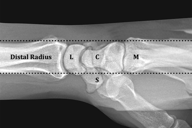
  • Normal distal radius(R), capitate C), lunate(L), scaphoid(S) and metacarpals(M) alignment on “true” lateral x-ray of the wrist. A “true” lateral x-ray of the wrist must be taken in neutral forearm rotation and neutral wrist deviation.
    Normal distal radius(R), capitate C), lunate(L), scaphoid(S) and metacarpals(M) alignment on “true” lateral x-ray of the wrist. A “true” lateral x-ray of the wrist must be taken in neutral forearm rotation and neutral wrist deviation.
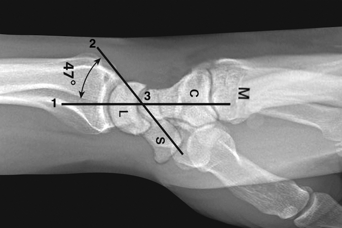
  • On a normal neutral rotation lateral, the horizontal axis of the radius, lunate, capitate, and metacarpals is a straight line (1).  A line (2) the longitudinal axis of the scaphoid crosses line (1) at point (3).  The average normal angle between these lines is 47° (range 30-60°).  Angles outside this range suggest carpal instability (DISI >60°; VISI <30°).
    On a normal neutral rotation lateral, the horizontal axis of the radius, lunate, capitate, and metacarpals is a straight line (1). A line (2) the longitudinal axis of the scaphoid crosses line (1) at point (3). The average normal angle between these lines is 47° (range 30-60°). Angles outside this range suggest carpal instability (DISI >60°; VISI <30°).
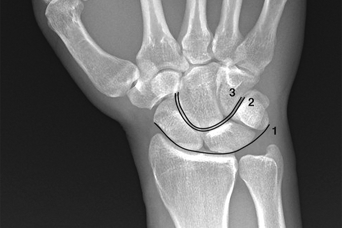
  • Gilula’s lines (ref 2) superimposed on a neutral deviation PA wrist x-ray. Arc 1 is a smooth arcing line paralleling the proximal articular surfaces of the triquetrum, lunate, and scaphoid. Arc 2 parallels the distal concave surfaces of the triquetrum, lunate, and scaphoid. Arc 3 parallels the smooth curved surface of the proximal hamate and capitate. When these smooth curved lines are irregular, disrupted, or step off it is indicative of a carpal instability or dislocation.
    Gilula’s lines (ref 2) superimposed on a neutral deviation PA wrist x-ray. Arc 1 is a smooth arcing line paralleling the proximal articular surfaces of the triquetrum, lunate, and scaphoid. Arc 2 parallels the distal concave surfaces of the triquetrum, lunate, and scaphoid. Arc 3 parallels the smooth curved surface of the proximal hamate and capitate. When these smooth curved lines are irregular, disrupted, or step off it is indicative of a carpal instability or dislocation.
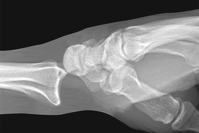
  • Diagrammatic lateral x-ray of a dorsal radiocarpal dislocation.  R-radius; L-lunate; C-capitate; Entire carpus is displaced dorsally.
    Diagrammatic lateral x-ray of a dorsal radiocarpal dislocation. R-radius; L-lunate; C-capitate; Entire carpus is displaced dorsally.
Symptoms
History of high energy severe wrist trauma
Wrist pain and swelling localized at the radiocarpal joint
Wrist deformity
Numbness and tingling in the hand
Typical History

The typical patient is a 31-year-old left-handed male contractor who injured himself on the job. The man was securing a beam of the second story about 8 feet above the ground level when he lost his balance and fell forward, landing on his outstretched right hand. The impact caused a severe shear force that was transmitted to the radiocarpal joint, and the result was a closed dislocation of this joint. Wrist pain, swelling and significant deformity were immediately noted. A co-worker splinted the wrist with a small piece of wood and an ace bandage.  The worker was then taken to a local emergency room.  An X-ray showed a dorsal radiocarpal dislocation.  A hand surgeon then reduced the dislocation under a local anesthetic block and IV sedation.  A traction X-ray was taken during the reduction.  After reduction a sugar tong splint was applied.  The traction X-ray showed a radial styloid fracture and ulnar translation of the carpus.  The patient was admitted.  The following day, a formal ORIF of the fracture was done and the dorsal and volar ligaments were surgically repaired.

Positive Tests, Exams or Signs
Work-up Options
Images (X-Ray, MRI, etc.)
Radiocarpal Dislocation
  • Lateral X-ray of a volar radial carpal dislocation in a 51 y.o. male involved in a MVA.  Note the associated mid-carpal gapping (arrow) suggesting some additional inter-carpal ligament damage.
    Lateral X-ray of a volar radial carpal dislocation in a 51 y.o. male involved in a MVA. Note the associated mid-carpal gapping (arrow) suggesting some additional inter-carpal ligament damage.
  • AP  X-ray of a volar radial carpal dislocation.  Note carpal bones (arrow) overlapping the distal radius.
    AP X-ray of a volar radial carpal dislocation. Note carpal bones (arrow) overlapping the distal radius.
Treatment Options
Treatment Goals
  • Reduce the dislocation
  • Analyze the radiocarpal joint’s stability
  • Perform appropriate ORIF of associated fractures and ligament repairs
  • Rehab the wrist to regain range of motion 
  • Preserve near normal wrist and hand function
Conservative
  • A standardized treatment protocol has not yet been established for radiocarpal dislocations, and the optimal management of these injuries is not known.3,5
  • Conservative treatment consisting of closed reduction and cast immobilization may be utilized for some radiocarpal dislocations, but most surgeons generally prefer surgical intervention, as casting only is associated with poor outcomes and higher rates of carpal instability.3-5
  • One situation in which conservative treatment may be indicated is for simple, closed radiocarpal dislocations with no associated intracarpal ligament injuries.
    • In these cases, the injury can be reduced closed, and if the radiocarpal joint remains stable without intracarpal damage after reduction, conservative treatment may be continued. Otherwise, surgery is indicated.9
Operative
  • Most surgeons prefer to manage radiocarpal dislocations surgically to reduce the risk of long-term carpal instability and other complications.3-5,7
  • Surgical treatment should be initially directed at stabilization and soft tissue coverage, and available options include closed reduction and percutaneous pinning (CRPP), open reduction and internal fixation (ORIF), primary ligamentous repair, lunoradial arthrodesis, or some combination of these interventions.7,8
  • ORIF
    • Agreement on the ideal surgical approach for radiocarpal dislocations has not yet been reached.5
    • After a dislocation with no intracarpal damage is reduced, the radiocarpal ligaments should be repaired through a volar approach and protected by radiocarpal K-wire for 2 months.11
    • Dislocations with intracarpal damage typically require anatomical reduction that is verified through a dorsal approach and stabilized by radial styloid pinning.11
    • Several authors advocate for both a volar and dorsal approach to treat volar dislocations, and this may be appropriate in the acute as well as the chronic setting.5
  • Arthrodesis
    • When initial fixation methods fail, radioscaphoid, radiolunate, or total wrist arthrodesis may be a necessary salvage procedure.7
  • Ligamentous reconstruction
    • Attenuated radioscaphocapitate ligament reconstruction with the brachioradialis tendon may be considered an alternative to wrist arthrodesis in certain cases but usually need the assistance of va hand therapist.7,12
Treatment Photos and Diagrams
Radiocarpal dislocation
  •  Volar radial carpal dislocation in a 51 y.o. male (insert) after closed reduction and before open ligament repair.
    Volar radial carpal dislocation in a 51 y.o. male (insert) after closed reduction and before open ligament repair.
  •  AP X-ray of volar radial carpal dislocation in a 51 y.o. male after closed reduction.
    AP X-ray of volar radial carpal dislocation in a 51 y.o. male after closed reduction.
Hand Therapy
  • Patients with closed radiocarpal joint dislocations that are reduced early may be able to exercise their wrist on their own.
  • However, patients with marked swelling and pain will need hand therapy to help reduce swelling and improve ROM and strength.
  • Surgically repaired complex radiocarpal dislocations, repaired ligaments, and unstable radiocarpal dislocations will definitely need hand therapy, custom splinting, and dynamic extension splints after initial splinting and casting for six weeks.
  • After surgery, immobilization with a long-arm cast should be initiated for 3-10 weeks, depending on the amount of time needed for the involved ligament(s) to heal.9
Complications
  • Stiffness
  • Pain
  • Residual deformity
  • Weakened grip
  • Loss of range of motion 
  • Compartment syndrome
  • Nerve injuries 
  • Vascular injuries
  • Carpal tunnel syndrome
  • Chronic carpal instability
  • Osteoarthritis - rates of post-traumatic osteoarthritis have ranged from 11-25% of cases.8
  • Ulnar translocation of the carpus is likely caused by insufficient ligamentous healing and is seen most commonly in dorsal dislocations with no or small radial styloid fractures .7,12,13
Outcomes
  • Although it is difficult to make direct comparisons between the two treatment approaches due to low incidence rates and lack of research, it appears that outcomes are superior and carry a lower risk for carpal instability in patients receiving surgery compared to conservative treatment. Early surgical intervention involving restoration of radiocarpal alignment and stability is therefore recommended to achieve the best functional outcome possible.3-5,8
  • In general, pure radiocarpal dislocations without associated bony injury have an inferior outcome to fracture-dislocations.2 Fractures heal better than ligaments.12,13
  • Anatomical restoration of the torn ligamentous structures and temporary stabilization of the radiocarpal joint offer the wrist the optimal chance for improved functional outcome and can lead to very good functional results, even if time since injury is extended by a few weeks.5
  • In a review of 23 cases of volar radiocarpal dislocation, long-term clinical results were disappointing, with reports of loss of strength and range of motion, volar radiocarpal subluxation, ulnar and/or volar carpus translation, and other complications being common.9
  • All patients with radiocarpal joint dislocations should be warned that the radiocarpal joint on the injured side will likely remain slightly larger than the opposite radiocarpal joint.
Key Educational Points
  • Open and complex radiocarpal joint dislocations require urgent surgical treatment.
  • Delayed and missed diagnoses of a radiocarpal dislocation can occur particularly if the joint is only partially dislocated (subluxated).  Therefore, a high index of clinical suspicion shouild be maintained with any high-energy wrist injuries. This is particularly necessary when a patient complains of severe pain and edema but initial radiographs are reported as normal.3
  • When a radiocarpal dislocation is purely ligamentous, or only a small volar articular rim fracture fragment is present, fixation is often challenging.5
  • Some radiocarpal dislocations are obvious on plain radiographs, but traction radiography may be needed to evaluate occult injuries.3
  • CT scan or MRI may be needed occasionally for better visualization of the injury and any associated lesions undetected on standard radiographs.3
  • Wrists that are already damaged by inflammatory arthritis (i.e. rheumatoid arthritis), congenital deformities or previous trauma may be especially susceptible to radiocarpal dislocation and/or ulnar translocation.12
  • Dorsal radiocarpal dislocations require disruption of the volar radiocarpal ligaments or a displaced radial styloid fracture where the styloid fragment is the proximal attachment (origin) of the volar radiocarpal ligaments.12,13
  • A radiocarpal doslocation with a simultaneous ulnar translation of the carpus requires a global ligamentous injury.  The volar radiocarpal ligaments and the dorsal capsular attachment to the distal radius must both be ruptured.12
  • Brachioradialis tendon reconstruction of the volar radiocarpal ligaments and/or the dorsal capsule attachement is a useful tool when dealing with a global radiocarpal dislocations.12,13
  • Radiocarpal dislocations that occur in patients with pre-existing RA may require a lunoradial arthrodesis to stabilize their wrist after this dislocation.12,13
References

New and Cited Articles

  1. Dunn, AW. Fractures and dislocations of the carpus. Surg Clin North Am 1972;52(6):1513-38. PMID: 4564432
  2. Ilyas, AM and Mudgal, CS. Radiocarpal fracture-dislocations. J Am Acad Orthop Surg 2008;16(11):647-55. PMID: 18978287
  3. Singisetti, K, Konstantinos, M and Middleton, A. Volar radiocarpal dislocation: case report and review of literature. Hand Surg 2011;16(2):173-5. PMID: 21548154
  4. Cowley, R and Yoon, A. Pure ligamentous volar radiocarpal dislocation: a case report and discussion. ANZ J Surg 2018;88(6):E554-E555. PMID: 27062672
  5. Hofmeister, EP, Fitzgerald, BT, Thompson, MA, et al. Surgical reconstruction of a late-presenting volar radiocarpal dislocation: a case report. Am J Orthop (Belle Mead NJ) 2008;37(2):96-9. PMID: 18401486
  6. Merrell G, Slade JF. Dislocations and ligament injuries in the digits. In: Wolfe, SW, Hotchkiss RN, Pederson WC, Kozin SH (eds): Green’s Operative Hand Surgery.  Philadelphia 2011: Elsevier Churchill Livingstone, pp. 291-332.
  7. Obafemi, A and Pensy, R. Palmar radiocarpal dislocation: a case report and novel treatment method. Hand (N Y) 2012;7(1):114-8. PMID: 23450977
  8. Yuan, BJ, Dennison, DG, Elhassan, BT, et al. Outcomes after radiocarpal dislocation: a retrospective review. Hand (N Y) 2015;10(3):367-73. PMID: 26330765
  9. Jardin, E, Pechin, C, Rey, PB, et al. Open volar radiocarpal dislocation with extensive dorsal ligament and extensor tendon damage: A case report and review of literature. Hand Surg Rehabil 2016;35(2):127-34. PMID: 27117127
  10. Moneim, MS, Bolger, JT and Omer, GE. Radiocarpal dislocation--classification and rationale for management. Clin Orthop Relat Res 1985;(192):199-209.PMID: 3967423
  11. Spiry, C, Bacle, G, Marteau, E, et al. Radiocarpal dislocations and fracture-dislocations: Injury types and long-term outcomes. Orthop Traumatol Surg Res 2018;104(2):261-266.PMID: 29428553
  12. Maschke SD, Means KR, Parks BG, Graham TJ.  A radiocarpal ligament reconstruction using brachioradialis for secondary ulnar translation of the carpus following radiocarpal dislocations: a cadaver study. J Hand Surg AM. 2010; 35A: 256-261.
  13. Dumontier C, Reckendorf MZ, Sautet, Lenoble E, Saffar P, Allieu Y.  Radiocarpal dislocations: classification and proposal for treatment a review of twenty-seven cases. J Bone J Surg 2001; 83A: 212-218.

Reviews

  1. Yuan, BJ, Dennison, DG, Elhassan, BT, et al. Outcomes after radiocarpal dislocation: a retrospective review. Hand (N Y) 2015;10(3):367-73. PMID: 26330765
  2. Mourikis A, Rebello G, Villafuerte J, et al. Radiocarpal dislocations: review of the literature with case presentations and a proposed treatment algorithm. Orthopedics2008;31(4):386-92. PMID: 18453177
  3. Dumontier C, Reckendorf MZ, Sautet, Lenoble E, Saffar P, Allieu Y.  Radiocarpal dislocations: classification and proposal for treatment a review of twenty-seven cases. J Bone J Surg 2001; 83A: 212-218.

Classic

  1. Reynolds IS. Dorsal radiocarpal dislocation. Injury1980;12(1):48-9. PMID: 7203624
  2. Gilula LA: Carpal Injuries: Analytic Approach and Case Exercises.  Am J Roentgenology 133:513, 1979.