MRI Distal Biceps Rupture
-

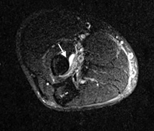
1. This MRI (T2) cross-sectional image at the level of the elbow shows the radius and the insertion site of the biceps tendon on the radius tuberosity (arrow). The ruptured biceps tendon is not present. Edema (white) secondary to the biceps rupture is evident at the tuberosity. The structures adjacent to the edema are the neurovascular bundle and surrounding fatty tissues.
-

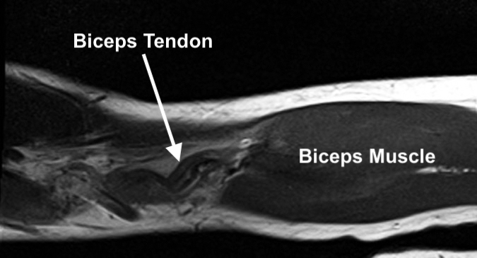
2. This MRI (T1) image of the biceps is a coronal view. The biceps muscle is visible, and the retracted curled up and ruptured biceps tendon is seen at the level of the elbow.
-

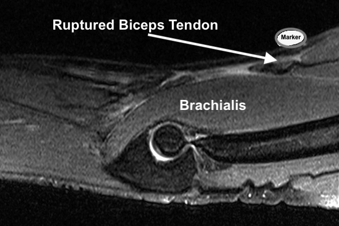
3. This MRI (T2) image of the elbow shows the humerus and olecranon with the brachialis muscle anterior to the humerus and elbow. In the proximal aspect of the image there is a marker on the skin (arrow) over the retracted ruptured biceps tendon. Fluid is noted in the elbow joint.
